Golf Ball Syndrom During Surrogacy Pregnancy
BlogGolf Ball syndrome during the pregnancy
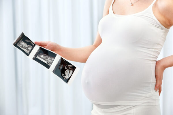
Intended parents are quite excited and at the same time very emotional during the pregnancy process of their surrogate mother. They are staying thousands of miles away from that special person who carries their baby all 9 month. After SM gets pregnant there are number of medical tests conducted on monthly basis to ensure smooth pregnancy process. Ultrasonography is one of the main scan to be conducted in order to check baby`s latest condition and development. I would like to talk about one of the issue which parents come across in the scan report and it make them worry to ready out this information. It is called Golf Ball syndrome.
Golf Ball is a small white spot, which can be seen in the fetus heart while doing a routine ultrasound scan. In medical terminology Golf Ball is called Echogenic Cardiac Foci. When Golf ball issue is identified in most of the cases it is considered harmless. This ball is a thickening of one of the cord which hold the heart valve. This kind of thickening does not have an effect on the heart functioning or its structure , unless no other heart defect are identified.
Most of the time echogenic foci is visible in the left ventricle of the heart. But sometimes in both ventricle it is detectable. Statistically during the pregnancy about 4 percent of babies may have this spot. Even though Echogenic foci is most of the time harmless, it could be related to heart defect of the baby too.
What measures need to be taken when echogenic cardiac foci is identified?
During the pregnancy of surrogate mother we perform 4 dimensional ultrasound scan and examine all the organs development in detail. Radiologist records a video on CD and parents are able to see the fetus movement and all the measurement of the organs are written in the chart. 4 D scan is recommended to be performed at pregnancy week 20-25. This is a time period when fetus organs have completed its development. Most of the time Golf ball is identified after II trimester of pregnancy. And sonographer has to evaluate how problematic it could be depend on its size, location. And if there is no other defect visible then radiologist will clarify for sure that echogenic foci does not cause any further trouble. Golf ball could be identified in one routine ultrasound scan but after 3-4 weeks period when ultrasound scan is repeated it could be disappeared itself.
From our side detailed examination is being performed by the professional doctors and relevant measures will be taken to make sure that echogenic foci will not cause any further complication for the fetus heart development.